Macular Degeneration
Macular Degeneration Treatment in Naples & Fort Myers, FL
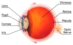
Macular Degeneration (AMD) is the leading cause of irreversible visual impairment and blindness among Americans 60 and older. More Americans are affected by AMD than by cataracts and glaucoma combined. There are two types of Macular Degeneration, Dry Macular Degeneration, and Neovascular, or Wet Macular Degeneration. In most cases, the dry type of macular degeneration usually progresses more slowly than the wet type. Approximately 90% of the cases of AMD are the dry type.
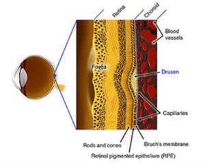
The retina is a very thin sheet of nerve cells that lines the inside of the eye. Macular Degeneration refers to a retinal disorder that affects the macula, or the center portion of the retina that is responsible for fine, detailed vision. Under the retina is a layer of retinal pigment epithelial cells (RPE), and under this is the choroid, a layer of tissue that is made up of blood vessels that bring essential nutrients and oxygen to the retina.
What is Dry Macular Degeneration?
There are many characteristics of dry AMD – some are very mild and do not affect the vision, some can cause moderate vision disturbances, and some can cause severe visual symptoms. Among these characteristics are Pigment Changes, Drusen, and Geographical Atrophy.
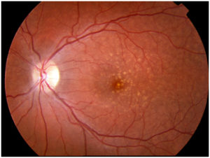 All cases of macular degeneration begin as the dry type. As the retina ages, the tissue in the macula begins to break down and pigment changes can occur. Pigment changes are small areas in the RPE layer where the cells can change color and clump together. Drusen are small yellow deposits in the retina that act like trash cans and collect waste products. They can multiply, progress and grow larger, and can overflow. This growth and overflow can cause blockage of nutrients to the retina and RPE cells. When RPE cells die, the overlying retina can begin to deteriorate. This is called atrophic macular degeneration.
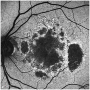 The advanced form of atrophic macular degeneration is referred to as Geographic Atrophy. Geographic atrophy is a disease affecting approximately ten million older Americans and causes blindness in over one million.
There is currently no effective treatment for geographic atrophy, as its cause is unknown.
According to some experts, the fact that no standard therapy exists may come from a lack of understanding of dry AMD disease. It is known that there are events that lead to the formation of drusen, but it is unknown whether that is due to inflammation, ischemia, genetic predisposition, or other factors. Risk factors for progression are known and appreciated but are just beginning to be understood. Most drugs being tested go straight to human testing instead of being tested in animals because atrophic AMD cannot be induced. This makes dry AMD a difficult disease to treat.
The fortunate news for the retinal community is that there are several drugs currently in various stages of development for the treatment of this common disorder. Many potential therapies with very different mechanisms of action are being investigated, a fact that increases the odds that a suitable therapy will be discovered.
Common symptoms of atrophic macular degeneration are decreased reading ability, especially in dim light, and difficulty in adapting to dim light and the dark. A vision test can sometimes reveal physical deterioration before symptoms occur. Early detection of atrophic AMD is important to check for signs of the more debilitating neovascular AMD.
To Learn More about Dry AMD, please visit the following websites:
- Fifteen-year Cumulative Incidence of Age-Related Macular Degeneration: the Beaver Dam Eye Study
- Healthy Lifestyles Related to Subsequent Prevalence of Age-Related Macular Degeneration
- Risk factors associated with age-related macular degeneration. A case-control study in the age-related eye disease study: Age-related Eye Disease Study Report Number 3
- The Age-Related Eye Disease Study: a clinical trial of zinc and antioxidants- Age-Related Eye Disease Study Report No. 2.
What is Wet Macular Degeneration?
Although only about 10 percent of all people with AMD have the wet form, it accounts for about 90 percent of all severe vision loss from the disease. As Dry AMD worsens, abnormal blood vessels behind the retina can start to grow under the macula, causing Wet AMD. These new blood vessels tend to be very fragile and they will often leak blood and fluid which can cause the loss
To learn more about Wet AMD, visit the following websites:
- Improved vision-related function after ranibizumab treatment of neovascular age-related macular degeneration: results of a randomized clinical trial
- Ranibizumab and Bevacizumab for neovascular age-related macular degeneration
- Intravitreal aflibercept (VEGF trap-eye) in wet age-related macular degeneration
What are the Symptoms of AMD?

- Decreased vision
- Blurred central vision
- Distortion of straight lines
- Dark or blank spots centrally
If Dry AMD changes to Wet AMD, symptoms may develop very quickly. The best way to detect if AMD is progressing is by checking the Amsler Grid each day. If the lines begin to become distorted, please contact Retina Consultants.
How is AMD Detected
The early and intermediate stages of AMD usually start without symptoms. Only a comprehensive dilated eye exam can detect AMD. The eye exam may include the following:
- Visual acuity test: The eye chart will measure how well you can see at a distance.
- Dilated eye exam: Eye drops will be placed in your eyes to widen or dilate the pupils. This gives the doctor a better view of the back of your eye. Using a special magnifying lens, your doctor then looks at your retina and optic nerve for signs of AMD and other eye problems.
- Fluorescein Angiogram: With this test, dye is injected into your arm. Pictures are taken as the dye passes through the blood vessels in your eye. This test allows your doctor to identify leaking blood vessels and recommend the best treatment option for you.
Video: Fluorescein Angiography
- Ocular Coherence Tomography: With this test, special pictures are taken to show a three-dimensional view of your macula. This test allows your doctor to determine if abnormal swelling or fluid is present.
- Amsler Grid: Your doctor also may ask you to look at an Amsler Grid. With AMD, changes in your central vision may cause the lines in the grid to blur, disappear, or appear distorted.
- Genetic Testing: Retina Consultants of Southwest Florida offers genetic testing to determine the presence of the genes associated with the development of AMD.
What are the Treatment Options
Currently, there is no cure for AMD, but there is hope.
With early diagnosis and proper treatment, the progression of AMD can often be delayed. The earlier AMD is detected, the better your chances are of keeping your vision.
For Dry AMD – There are currently no medications available to treat Dry AMD, but certain lifestyle changes and vitamin therapies may be helpful to slow the progression of the disease. Your Retina Consultants physician can discuss these with you in more detail.
For Wet AMD – Retina Consultants of Southwest Florida offers multiple treatment options to stop further vision loss in patients with Wet AMD:
- VEGF Inhibitors: The most common option to slow the progression of wet AMD is to inject medication into the eye. With wet AMD, abnormally high levels of vascular endothelial growth factor (VEGF) are present in the affected eye. This substance causes the growth of new abnormal blood vessels, which tend to be fragile and leak blood or other fluid. The medication injected into the eye can block the effects of VEGF. If you and your doctor choose this treatment, you will likely need multiple injections, often monthly. Before each injection, your doctor will numb your eye and clean it with antiseptics to prevent infection. There are currently multiple VEGF inhibiting treatment options available to patients with neovascular AMD.
- Macugen (pegaptanib) was the first FDA approved VEGF inhibitor and was available to patients in 2004. It had a higher success rate than photodynamic therapy. It is not commonly used today.
- Lucentis (ranibizumab) was FDA approved for use in patients with neovascular AMD in 2006 and has a much higher success rate than Macugen. It is one of three commonly used VEGF inhibitors today. It is manufactured by Genentech. For more information, go to www.lucentis.com.
- Avastin (bevacizumab), also manufactured by Genentech, is FDA approved for use in patients with colorectal cancer. Since it is very similar to Lucentis, it is commonly used in patients with neovascular AMD in an ‘off-label’ manner. Off-label means that the drug was FDA approved for a different condition than what it is being used for. The National Institute of Health recently sponsored a study, the CATT study, comparing Lucentis to Avastin in patients with neovascular AMD and found that the effects of each drug are comparable. For more information, go to www.avastin.com.
- Eylea (aflibercept) is the most recent FDA approved VEGF inhibitor. It is manufactured by Regeneron Pharmaceuticals and is another treatment option for patients with neovascular AMD. Studies are currently being conducted to compare Eylea with Lucentis and Avastin. For more information go to www.eylea.com.
Video: Eye Injection for Wet AMD
- Photodynamic Therapy (PDT) This less common treatment involves using a low-intensity laser for select areas of the retina. A light-sensitive drug called verteporfin will be injected into a vein in your arm or hand. This drug travels through the blood vessels in your body, including any new, abnormal blood vessels in your eye. Your doctor then shines a laser beam into your eye to activate the drug in the blood vessels. Once activated, the drug destroys the new blood vessels and slows the rate of vision loss. You will have to protect all of your skin from sun exposure for three days following this treatment.
Video: Photodynamic Therapy
- Laser treatment: Even though it is less common than other treatments, doctors sometimes treat certain cases of Wet AMD with laser treatment. It involves aiming a beam of light at the new blood vessels in your eyes to destroy them.
Video: Laser Treatment for Wet AMD
What Are The Causes And Risk Factors Of AMD?
- Age – People over the age of 50 are at greater risk than any other age groups, and the risk increases with age.
- Genetics – Recent studies have shown that there is a strong genetic component causing AMD. Certain genetic mutations may be responsible for AMD. Several other Environmental and Behavioral risk factors have been identified as having a possible relationship to AMD.
- Race – Caucasians are much more likely to get AMD than people of African descent.
- Family History – Those with a family history of AMD are more likely to develop AMD.
- Smoking – Smoking and second-hand smoke can drastically impact your chances of worsening AMD.
Other possible risk factors may include:
- Poor Diet
- Heart disease
- Sun exposure
- Light colored skin and eyes
- Farsightedness and Nearsightedness
- Cataract surgery
- High blood cholesterol
- Estrogen and early menopause
If your eye doctor recommends the AREDS vitamins, please consult your general medical doctor, as high dose vitamins can sometimes be harmful with certain medical conditions.
If you have wet AMD, it is important not to delay evaluations and treatment. It is very common to experience frequent flare-ups of Wet AMD after successful treatment. You will need to have frequent eye examinations as directed by your doctor.
People with both forms of AMD should monitor their eyesight each day by checking the Amsler grid. You should notify your doctor immediately if you notice any changes in your vision on the Amsler Grid.
Studies have shown that people who eat a diet rich in green, leafy vegetables and fish have a lower risk of developing AMD.
While there is no definitive proof that changing your diet will reduce your risk of developing AMD or its progression, Retina Consultants of Southwest Florida recommends eating a healthy diet, exercise, avoid smoking, and see your healthcare professional regularly.
Amsler Grid Self Test
The Amsler Grid Self Test can be an important indicator of diseases of the retina.
It is not a substitute for regularly scheduled eye exams or tests.
Amsler Grid Self Test
Video: Amsler Grid
Making the Most of Your Remaining Vision
Early detection and treatment may reduce the loss of vision from macular degeneration. However, if some loss of vision should occur, it doesn’t have to rob you of life’s simplest pleasures if you learn how to use your remaining eyesight to see your best. Low vision aids, special lenses, or electronic systems and training can maximize your ability to read and perform other activities.
The Low Vision Rehabilitation Center of Retina Consultants of Southwest Florida can give you more information about the training and devices available.
